
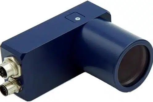
About Neuromorphic camera:
- A neuromorphic camera mimics the way the human retina converts light into electrical impulses.
How does it work?
- In a typical camera, each pixel captures the intensity of light falling on it for the entire exposure time the camera focuses on the object. All these pixels are pooled together to reconstruct an image of the object.
- In neuromorphic cameras, each pixel operates independently and asynchronously, generating events or spikes only when there is a change in the intensity of light falling on that pixel.
- This generates sparse and lower amounts of data compared to traditional cameras, which capture every pixel value at a fixed rate, regardless of whether there is any change in the scene.
- This allows a neuromorphic camera to “sample” the environment with much higher temporal resolution because it is not limited by a frame rate like normal cameras and also performs background suppression.
- Neuromorphic cameras have a very high dynamic range ( >120 dB) which means they can be used in different conditions ranging
- from a very low-light environment to very high-light conditions.
What is meant by diffraction limit in Optical Microscopy?
- The resolution of a microscope is proportional to the size of its objective and inversely proportional to the wavelength of light being observed.
- In 1873 the German physicist Ernst Abbe discovered how microscopes were limited by the diffraction of light. He revealed that the resolution of a microscope is not controlled by the instrument’s quality but by the wavelength of light used and the aperture of its optics.
- Due to this phenomenon, a microscope cannot resolve two objects located closer than λ/2NA, where λ is the wavelength of light and NA is the numerical aperture of the imaging lens. This is known as the diffraction limit.
- Thus diffraction limits the ability of the microscope to distinguish between two objects divided by a lateral distance of less than half the wavelength of light used to image the sample.
Q1) What is meant by temporal resolution?
Temporal resolution is the time to take the multiple measurements of the cross-section and then reconstruct the image. This time is important for process tomography systems, as changes can often occur within the system in a space of time that is shorter than the one required to record the measurements.
Source: IISc shows how neuromorphic camera, machine learning aid nanoscopic imaging


Last updated on July, 2025
→ UPSC Notification 2025 was released on 22nd January 2025.
→ UPSC Prelims Result 2025 is out now for the CSE held on 25 May 2025.
→ UPSC Prelims Question Paper 2025 and Unofficial Prelims Answer Key 2025 are available now.
→ UPSC Calendar 2026 is released on 15th May, 2025.
→ The UPSC Vacancy 2025 were released 1129, out of which 979 were for UPSC CSE and remaining 150 are for UPSC IFoS.
→ UPSC Mains 2025 will be conducted on 22nd August 2025.
→ UPSC Prelims 2026 will be conducted on 24th May, 2026 & UPSC Mains 2026 will be conducted on 21st August 2026.
→ The UPSC Selection Process is of 3 stages-Prelims, Mains and Interview.
→ UPSC Result 2024 is released with latest UPSC Marksheet 2024. Check Now!
→ UPSC Toppers List 2024 is released now. Shakti Dubey is UPSC AIR 1 2024 Topper.
→ Also check Best IAS Coaching in Delhi